3D Images of Lower Molars
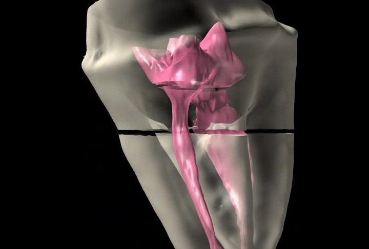
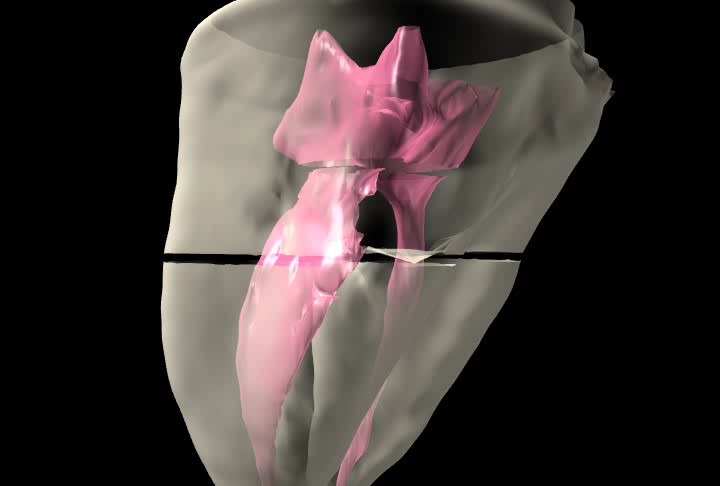

The images that my automatic data aquisition robot, "charlie" yields shows lower molars with the very same kind of inter-canal anatomy that my PVS impressions of root canal anatomy showed.
It also revealed a helical ribbon shaped distal canal in this molar.
I like viewing 3D images because they provide a guideline that the final fill should approximate (in a larger more shaped way).
posted by Scott Perkins at 11:58 AM
![]()
![]()


0 Comments:
Post a Comment
<< Home